El índice anchura del asta frontal respecto a la distancia intercaudados decrece. Lo normal es entre 2.2 y 2.6.
El índice anchura del asta frontal respecto a la distancia intercaudados decrece. Lo normal es entre 2.2 y 2.6.


 |
| T2*-weighted (a and b), T2-weighted (c and d), and FLAIR (e and f) images of the medial temporal lobe obtained at 1.5 T (a, c, and e) and 7 T (b, d, and f), illustrating the strikingly improved resolution that high-field MRI offers. Reprinted from Theysohn et al.: The human hippocampus at 7 T-in vivo MRI, Hippocampus 19:1–7, 2009, copyright 2008, Wiley-Liss, Inc. |
 |
| Five AD hippocampal specimens (A1–A5) and one normal control (N4) are shown. Note the signal voids in AD specimens along the hippocampus compared with the lack of such signal voids in the normal control. The border between field CA1 and the subiculum is indicated by the white line derived from coregistered acetylcholine, myelin, and Nissl staining. The variability in their locations relative to the medial aspect of the hippocampal body illustrates the challenges inherent in in vivo imaging studies of hippocampal subregions. Reprinted from Neurobiology of Aging, vol 36, Zeineh M, Chen Y, Kitzler HH, Hammond R, Vogel H, Rutt BK, “Activated iron-containing microglia in the human hippocampus identified by magnetic resonance imaging in Alzheimer’s disease,” pp 2483–2500, 2015, with permission from Elsevier. |
 |
| 7-T FLAIR MRI in the (left to right) transverse (left), sagittal (center), and coronal (right) views. The arrow is pointing to a microinfarct. Reprinted with permission from van Rooden S, Goos JD, van Opstal AM, Versluis MJ, Webb AG, Blauw GJ, et al: Increased number of microinfarcts in Alzheimer disease at 7-T MR imaging. Radiology 270:205–211, 2014. |
 |
| A. Paciente con Atrofia Multisistema, presenta una morfología del bulbo olfatorio normal. B. Paciente con Parkinson, con reducción del volumen del bulbo. |
 |
| Espectro somatotópico de lesiones en distintas afectaciones del nervio ciático demostradas por RM |
 |
| 7T T2*-weighted axial images from (A) an MS patient using the Axial1 protocol (in-plane resolution: 215 × 286 μm). Images demonstrate multiple foci of abnormality involving the deep WM structures, as well as the GM, WM, and lesion contrast achieved using this methodology, and (B) an MS patient with multiple lesions using the Axial2 protocol (in plane resolution: 195 × 260 μm), along with an enlarged image of highlighted selection which demonstrates sub-cortical WM lesions (arrows). |
 |
Neuroimaging in patients with an immunotherapy-responsive cognitive disorder.
Magnetic resonance imaging: yellow arrows indicate areas of abnormality on fluid-attenuated inversion recovery (FLAIR). A, 36-year-old woman (patient 10 in Appendix 1) had fluctuating memory problems and was seropositive for glutamic acid decarboxylase-65 autoantibody. Bilateral hippocampal axial FLAIR abnormality, shown in A1, almost completely resolved after treatment with intravenous (IV) methylprednisolone (A2). B, 51-year-old woman (patient 20 in Appendix 1) had subacute fluctuating memory problems, multifocal neurologic examination findings, and evidence of autoimmunity (IgM antiphospholipid antibody). Symmetric confluent T2 signal abnormality in the white matter of both hemispheres (B1) decreased after treatment with IV methylprednisolone (B2). C, 60-year-old man (patient 41 in Appendix 1) had memory, language, and gait problems and was seropositive for both striational and glutamic acid decarboxylase-65 antibodies. Axial T1 magnetic resonance imaging with contrast demonstrated periventricular vessel enhancement (C1) and resolution after treatment (C2). D, 53-year-old woman (patient 29 in Appendix 1) had memory loss, hallucinations, and subsequent seizure; cerebrospinal fluid protein was elevated (>100 mg/dL), and she was seropositive for thyroid peroxidase antibodies and neuronal and muscle acetylcholine receptor antibodies. Axial FLAIR images show diffusely increased T2 signal in the midbrain (D1), which improved after treatment with IV methylprednisolone (D2). Multiple myeloma was diagnosed 18 months after neurologic presentation.
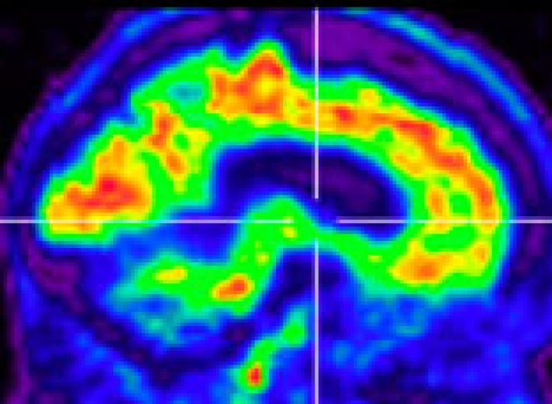
Positron emission tomographic imaging: Brain reconstructions (brighter color represents regions of hypometabolism) in a 58-year-old man (patient 21 in Appendix 1) who presented with personality change and memory problems and had elevated cerebrospinal fluid protein (>100 mg/dL). Hypometabolism, predominantly frontal and temporal (E1), improved after treatment with IV methylprednisolone (E2).
Single-photon emission computed tomographic brain imaging: Brain neuroimaging in a 35-year-old man (patient 22 in Appendix 1) who presented with vertigo and memory problems, had multiple coexisting autoimmune conditions, and was seropositive for muscle acetylcholine receptor and striational antibodies. Diffuse decrease in uptake in frontotemporoparietal regions (F1) was markedly improved globally after treatment with IV methylprednisolone (F2).
|
 |
Features of atypical anti-NMDA receptor encephalitis MRI: (A) Initial contrast-enhancing lesion; (B) longitudinally extensive transverse myelitis; (C) continued development of contrast-enhancing lesions; (D) retrochiasmatic optic neuritis; (E, F) continued accumulation of T2/fluid-attenuated inversion recovery (FLAIR) hyperintense lesion burden, with sagittal FLAIR hyperintensities reminiscent of Dawson's fingers (E). Brain biopsy (from contrast-enhancing frontal lobe lesion): (G) perivascular infiltrate with associated reactive microgliosis; (H) widespread parenchymal destruction mediated by infiltrative lymphocytes and macrophages without selective demyelination; (I) prominent mixed perivascular infiltrate (macrophages, T- and B-lymphocytes with uncommon neutrophils and rare eosinophils). Western blot: (J) Western blot depicting the presence of several additional serum-derived autoantibodies reactive against cerebellar protein extract from control human brain. |
 |
21.1-Tesla MRI on postmortem brain sections of the hippocampus Fixed postmortem samples were washed in phosphate-buffered saline and immersed in Fluorinert (3M, Corp). Utilizing a 21.1-T magnet (Bruker Avance console and Micro2.5 gradients) and 33-mm birdcage coil, 3-dimensional 1H fast low angle shot (FLASH) scans (echo time/repetition time = 12/50 msec) were acquired in 3-dimensional at 50-μm isotropic resolution over 4.3 hours at 14°C. (A) Normal hippocampal and (B) sclerotic sections. |